Brain shrinkage found to be worse, faster in pediatric Huntington’s
Pediatric-onset disease more severe than adult-onset, imaging study shows

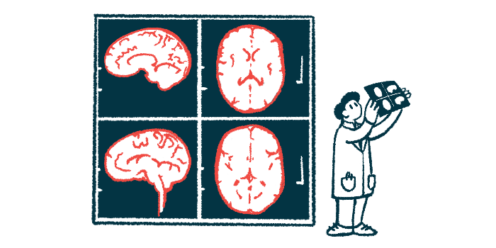
Pediatric-onset Huntington’s disease is more severe than its adult-onset form, with faster and more extensive shrinkage of brain regions involved in movement, an imaging study has found.
Atrophy, or a loss in brain volume, “was remarkable and more severe” in patients with pediatric-onset disease compared with adult-onset Huntington’s, the researchers reported.
Further, the team determined that changes in brain activity mirror clinical progression, with some changes possibly occurring even before the brain begins to shrink. This may predict how severe symptoms will be, according to the researchers, who noted that the identification of imaging biomarkers of brain activity and structure may help doctors track the disease and develop better treatments.
The researchers noted that their study was limited by its small size, but suggested that imaging scans “may represent a valuable tool for monitoring [pediatric-onset Huntington’s disease] changes over time.”
The study, “Pediatric Huntington Disease Brains Have Distinct Morphologic and Metabolic Traits: the RAREST-JHD Study,” was published as a brief report in the journal Movement Disorders Clinical Practice by researchers in Italy.
Using imaging scans to track brain shrinkage in patients
Regardless of its type, Huntington’s disease is caused by excessive repeats of three DNA building blocks — C, A, and G — in the HTT gene. Most often, symptoms of Huntington’s begin in adulthood, which is known as the adult-onset form of the disease. However, in as many as 10% of patients, symptoms manifest before age 21; this is known as juvenile-onset Huntington’s.
More rarely, symptoms begin even earlier, before age 18. This form of the disease, called pediatric-onset Huntington’s, or POHD, is linked to more than 55-60 CAG repeats.
Unlike adult-onset Huntington’s, which is characterized by involuntary, jerky movements known as chorea, the pediatric-onset form mainly manifests as slowed movements, called bradykinesia, muscle stiffness or rigidity, and abnormal postures, known as dystonia.
Previous research also showed that pediatric-onset disease differs from the adult-onset form in terms of brain metabolism, function, and degeneration, emphasizing the presence of different biological mechanisms.
According to the researchers, pediatric-onset Huntington’s “may therefore require a different treatment approach and the identification of distinct biomarkers for monitoring disease progression.”
To that end, “imaging studies integrating glucose [blood sugar] metabolism and regional brain volumes could identify such POHD-specific biomarkers for use in [Huntington’s disease] trials,” the team wrote.
With this in mind, the researchers combined two imaging techniques — positron emission tomography (PET) and magnetic resonance imaging (MRI) — to compare brain activity and structure in patients with pediatric-onset versus adult-onset Huntington’s.
The study, dubbed RAREST-JHD, included five adults with pediatric-onset Huntington’s and 14 with the adult-onset form who were followed for up to three years. The mean follow-up duration was slightly shorter than 1.5 years.
Those with pediatric-onset Huntington’s had significantly more CAG repeats (60 vs. 43), were significantly younger (mean age 24.2 vs. 45.1 years), and had their first symptoms at a significantly younger age (16 vs. 42.5 years).
Faster brain volume loss seen in pediatric-onset Huntington’s patients
At the study’s start, adults with pediatric-onset Huntington’s had significantly smaller structures of the striatum, a brain region involved in movement and selectively affected by Huntington’s. The patients with POHD also had a significantly thinner cortex, which is the outer layer of the brain, relative to those with adult-onset disease.
Over time, the pediatric-onset Huntington’s patients also lost brain volume faster, with the caudate, a part of the striatum, showing a reduction of as much as 30%. Cortical thickness showed a reduction of up to 15%. Also, ventricular spaces, or fluid-filled cavities in the brain, were enlarged by 30% in POHD patients, compared with 10% in those with adult-onset disease.
“Although previous studies have correlated longitudinal striatal volume loss with clinical progression, ours is the first to show that both baseline and longitudinal changes, in striatal and nonstriatal regions, may explain clinical differences between POHD and AOHD [adult-onset Huntington’s disease],” the team wrote.
Consistent with earlier findings of impaired glucose transport in the brain of pediatric-onset Huntington’s patients, striatal glucose metabolism, which indicates nerve cell activity in the striatum, was significantly lower in pediatric-onset versus adult-onset Huntington’s patients at the study’s start.
[Our study] is the first to show that both baseline [from the study’s start] and longitudinal changes, in [varying brain] regions, may explain clinical differences between POHD [pediatric-onset Huntington’s disease] and AOHD [adult-onset Huntington’s disease].
Over follow-up, glucose metabolism in the POHD group was significantly reduced in some brain regions and increased in others, while those with AOHD generally experienced widespread reductions in glucose metabolism.
“Alterations in glucose metabolism were highly individualized, with greater annual changes in metabolism linked to more severe worsening in annualized dystonia, parkinsonism [a measure of rigidity and slowness], and independence scores,” the team wrote.
The researchers added that “the regional and dynamic variability in cortical dysfunction that precedes cortical [shrinkage] is potentially a precursor of clinical manifestation severity in POHD.”
No significant differences in terms of clinical severity scores were seen at the study’s start. However, the POHD group had more parkinsonism, and this worsened over time. Symptoms like dystonia also worsened over time, limiting functional independence.
Altogether, these findings “provide further evidence that POHD is a distinct disease from AOHD,” the researchers wrote.
The study also showed PET/MRI is a valuable tool for studying pediatric-onset Huntington’s. It helped identify specific biomarkers such as striatum shrinkage and changes in glucose uptake, which may be used to track disease progression, according to the researchers.


