
Scans show brain changes decades before Huntington’s onset
Study suggests brain connections evolve differently for those carrying mutation
Written by |
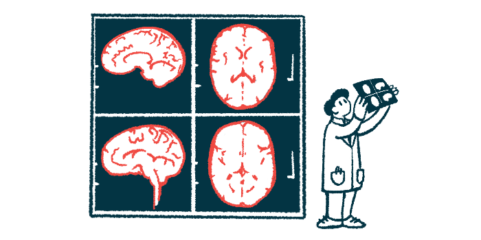
Differences in brain connections for people who carry a mutation known to cause Huntington’s disease are detectable decades before the onset of motor symptoms, a study showed.
The findings suggested that the ways these connections evolve over time is altered in Huntington’s mutation carriers.
While the study was limited to a relatively small number of participants, the data add to a growing body of evidence that changes in brain function can be seen years before symptoms and “highlight the potential of [brain connection] measures as sensitive biomarkers for [Huntington’s mutation-carrying] individuals before clinical motor diagnosis,” the researchers wrote.
“Identifying these early markers is essential for future clinical trials aimed at detecting treatment effects and preserving long-term quality of life,” they said.
The study, “Early Longitudinal Brain Network Changes in Huntington’s Disease Before Clinical Motor Onset,” was published in Movement Disorders.
Huntington’s is a genetic disorder that causes damage to certain regions of the brain, leading to Huntington’s symptoms including motor problems and cognitive issues. Huntington’s-causing mutations are present from the moment of conception, but most people don’t develop overt symptoms until well into adulthood.

Analyzing MRI data
The researchers wanted to better understand what’s happening in the brains of adults who carry a Huntington’s-causing mutation but who are not yet experiencing symptoms.
To do so, they analyzed MRI data from participants in the Huntington’s Disease Young Adult Study, an ongoing research effort to better understand disease biology prior to symptom onset. Participants are estimated to be approximately 24 years away from the first onset of motor symptoms.
The analysis looked at data on 43 Huntington’s mutation carriers and 28 control participants who did not have a mutation and were closely matched for age, sex, and interval between MRI scans. Participants underwent two MRI scans nearly five years apart.
Using the imaging data, the researchers looked at patterns of functional connectivity (FC), or how strongly different brain regions are connected. The team focused on the caudate and putamen, two brain regions that are heavily affected by Huntington’s.
Results showed several significant differences between the mutation carriers and controls. For example, carriers tended to have reduced FC between the putamen and other regions including the cerebellum and brainstem, both of which are important for controlling movement and coordination. This reduction in brain connectivity suggests that subtle brain damage is already occurring years before symptom onset.
“These early FC changes may represent a preclinical biomarker of impaired motor system integration,” the scientists wrote.
At the same time, mutation carriers had significantly greater FC between the putamen and the precuneus, a brain region involved in high-level cognition, visuospatial processing, and aspects of consciousness. The researchers said that this increase probably reflects healthy parts of the brain attempting to compensate for damage elsewhere.
“Our findings suggest that the increased FC observed in [Huntington’s mutation carriers] may represent either a compensatory recruitment of higher-order cognitive networks to offset early motor system changes,” the researchers wrote. “These patterns might represent adaptive reorganization aimed at preserving cognitive and motor function during the earliest stages of [Huntington’s disease].”
In both mutation carriers and controls, FC patterns changed over time. However, some FC trajectories differed significantly between groups, with mutation carriers showing greater FC reductions across several brain regions.
“Whether the observed FC differences reflect altered neurodevelopment, very early neurodegeneration, or their interaction remains unclear,” the researchers wrote, adding that long-term studies “are necessary to disentangle these possibilities.”
“Until then, interpretations … should be made with caution,” they said.



Leave a comment
Fill in the required fields to post. Your email address will not be published.