
Imaging technique may track brain cell damage in Huntington’s disease
Study: Method also shows promise for testing new therapies
Written by |
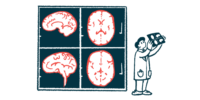
Soma and Neurite Density Imaging (SANDI), an imaging technique that uses MRI scans to capture microstructural features of the brain, may be a useful tool for tracking the progression of Huntington’s disease, a study suggests.
Huntington’s patients’ SANDI data, together with age, could explain up to 63% of shrinkage (atrophy) in the striatum, a brain region that’s profoundly affected by Huntington’s.
“SANDI shows significant promise for tracking Huntington’s disease and testing whether new therapies protect brain cells, and could also be applied to more common neurodegenerative conditions such as Alzheimer’s and Parkinson’s disease,” Claudia Metzler-Baddeley, the study’s senior author at the Cardiff University Brain Research Imaging Center in the U.K., said in a press release from eLife. “Before that can happen, our findings need to be confirmed in larger, long-term studies, and we hope our work will provide a useful framework for those studies.”
The study, “In vivo mapping of striatal neurodegeneration in Huntington’s disease with Soma and Neurite Density Imaging,” was published in eLife.

Tools that accurately detect striatum damage needed
Huntington’s is caused by mutations in the HTT gene, which result in the production of an abnormal huntingtin protein that is prone to forming toxic clumps in brain cells. Damage to the brain in people with Huntington’s typically starts in the striatum, which helps regulate movement, cognitive function, and emotion, and then spreads to other parts of the brain.
There is currently no treatment that has been proven to slow the development or progression of Huntington’s, but many are in development. To help determine their efficacy, there is a need for tools that can accurately detect striatum damage.
This is traditionally done by using standard MRI scans to measure the volume of different brain regions, but this gives limited insight into the underlying nerve cell damage.
“The recent surge in potential disease-modifying targets has generated a demand for surrogate outcome measures that are sensitive to [Huntington’s nerve damage] and allow a mechanistic assessment of therapeutic effects on striatal neurodegeneration in a timely manner,” the researchers wrote.
SANDI is a type of diffusion MRI analysis that uses mathematical formulae to estimate how much of the brain tissue consists of nerve cells’ rounded bodies (soma), their branching extensions (neurites), and the space between cells. Unlike standard MRI, diffusion MRI provides information about microscopic tissue structure by detecting the movements of water molecules in and around cells.

Huntington’s patients had fewer cell bodies, imaging shows
In this study, Metzler-Baddeley and colleagues used SANDI to analyze diffusion MRI data from 56 people with Huntington’s and 57 people without any neurological disorder.
They looked at two brain regions: the basal ganglia, which includes the striatum, and the thalami, which usually remains intact until Huntington’s disease is quite advanced.
“We wanted to test how SANDI performs in characterising Huntington’s disease-related abnormalities in the basal ganglia and thalami, and examine associations between SANDI indices, volumetric measurements, and motor performance,” Metzler-Baddeley said. “This is the first time, to our knowledge, that SANDI has been applied in Huntington’s disease, an approach with potential translatability to other neurodegenerative diseases.”
SANDI showed that people with Huntington’s had significantly lower soma density (fewer cell bodies) and significantly more space between cells across basal ganglia structures relative to healthy controls. At the same time, the nerve cell bodies in the striatum of Huntington’s patients were significantly larger than those in healthy volunteers.
Our findings highlight the potential of SANDI-derived metrics as future markers for tracking disease progression and assessing therapeutic efficacy in [Huntington’s].
As expected, no significant differences in SANDI metrics of the thalami were found between patients and healthy volunteers.
“This pattern of macro- and microstructural differences in the [basal ganglia] aligns with previous reports in premanifest and early manifest HD stages,” the researchers wrote. Premanifest refers to a disease stage where there are no overt motor symptoms yet.
Statistical models showed that SANDI metrics, together with age, could explain up to 63% of striatum atrophy in Huntington’s patients.
In patients, SANDI measurements also showed significant associations with measures of motor function and a score that estimates lifetime exposure to mutant huntingtin protein.
“Our findings highlight the potential of SANDI-derived metrics as future markers for tracking disease progression and assessing therapeutic efficacy in [Huntington’s],” the researchers wrote. “The observed associations between SANDI metrics, motor performance, and disease burden underscore their relevance for evaluating the neural effects of emerging disease-modifying treatments.”
The team noted, however, that more research is needed to validate the findings.




Michael Brunetti
That is awesome to know how is everything going with the research for everything? I have a Huntington. I’m not symptomatic right now, but I’m older. I’m 43 years old. We had my son through IVF and his Huntington free. he is nine years old. Everything is going good for us.
Jessica Morgan
Parkinson’s disease is such a difficult condition, and when my husband was diagnosed, it was heartbreaking for our whole family. We wanted to find additional support for him, so our family doctor recommended that we look into NaturePath Clinic and its herbal program.From the beginning, we appreciated the care, kindness, and support we received. Every family’s experience is different, but we are grateful for the encouragement and support NaturePath has provided to our family. ❤️If you or someone you love is looking for additional support, you can learn more about NaturePath by calling or contacting them on WhatsApp: +1 678 276 0310
Chris Pryor
I tested positive for Huntington corea age 50 years old with a CAG of 43 the same as my older brother who died of Huntington corea age 65 years old because Huntington Corea affect the brain I have not slept at all for 3 years and am exhausted and use a wheel chair inside and outside the care home I have problems with eating and drinking and use a straw to drink and my food is all mashed up I have corea in my hands and use plastic grips for eating with I have 2 children who both teasted negative for Huntington corea age 18 years old I have lived in Germany for 30 years I am 65 years old and live in a care home for 2 years in Jena Germany and my daughter and son both live in Jena Germany Chris Pryor