
Huntington’s Research Likely to Benefit from Device Aiding Studies of Neurological Diseases in a Living Animal
Written by |
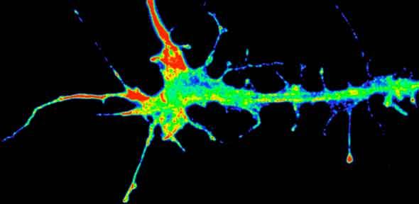
Credit: K-M. Leung
Researchers from the École Polytechnique Fédérale de Lausanne (EPFL), Switzerland, led by Dr. Matteo Cornaglia, PhD, have developed a new experimental device that can assist scientists in the analysis of molecular pathway studies related to complex human dysfunctions, such as neurodegenerative diseases like Huntington’s (HD). The device is intended for use with the experimental animal model Caenorhabditis elegans, commonly known as the nematode.
Nematodes are experimental models for studying numerous human diseases, but they have to be handled with care due to their delicate tissue structure. In an effort to make the use of this animal model in experimental protocols easier, the researchers created a new technology that allowed for the manipulation of nematodes within an aluminum framed 1-layered “chip” specifically designed for a balanced temperature distribution over its whole three-dimensional (3-D) geometry.
The chip is unique in that it does not require a valving system to distribute fluids, but instead uses passive hydrodynamics to properly distribute fluids throughout the structure. A hole in the center of the chip allowed for light transmission through which the researchers can observe the nematodes and take images using transmission microscopy.
In an EPFL press release explaining the significance of this chip, Dr. Laurent Mouchiroud, Nestle Chair in Energy Metabolism and study co-author, said, “Unlike conventional cultures in petri dishes, this device lets us monitor individual worms rather than a population of them.”
Using this new chip technology, scientists studying the formation of protein aggregates that are linked to Huntington’s disease may now observe the formation of these aggregates as the clusters develop, for the same worm can now easily be photographed several times.
“This is totally new, and it will help us learn more not only about how these aggregates (protein) grow, but also about the tissue in which they form. In addition, we have already been able to test and observe the effect of certain drugs on how the clusters form,” Dr. Mouchiroud said.
The development and utilization of this technology is described in the journal Molecular Neurodegeneration, in a study titled “Automated longitudinal monitoring of in vivo protein aggregation in neurodegenerative disease C. elegant models.”


Leave a comment
Fill in the required fields to post. Your email address will not be published.