
Disease-associated Molecular Fingerprint Identified in the Blood of Huntington’s Patients Using Light
Written by |
Raman spectroscopy, an analysis technique that uses light, identified a specific molecular fingerprint in the blood of people with Huntington’s disease, a study has found.
This technique may be useful as a non-invasive diagnostic tool to track disease progression and as a marker to measure responses to therapies in clinical trials.
The study, “Serum Raman spectroscopy as a diagnostic tool in patients with Huntington’s disease,” was published in the journal Chemical Science.
Huntington’s disease is caused by a repeating set of nucleotides, called a CAG repeat, in the HTT gene. The CAG repeat is present in healthy cells, but in the case of Huntington’s, it undergoes an even greater expansion, leading to progressive loss of neurons in the brain, causing involuntary movements and cognitive decline.
Huntington’s is currently diagnosed by examining family medical history, identifying changes in motor functioning, and cognitive and behavioral changes.
Brain imaging scans such as magnetic resonance imaging (MRI) also may be used to identify disease-associated changes in the brain. A definitive diagnosis eventually can be made by a genetic test to identify changes to the HTT gene.
Despite all these diagnostic tools, there are no accurate ways to mark the onset of Huntington’s disease.
Another alternative approach would be to find molecules in the blood that indicate the presence of the disease, known as biomarkers. However, biomarkers that have been identified, to date, are associated with other neurodegenerative diseases, such as Alzheimer’s, or aging.
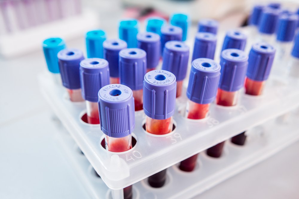
To identify biomarkers in blood that are specific to Huntington’s disease and disease progression, a team of researchers based at the University of Cambridge in the United Kingdom used a technique called Raman spectroscopy (RS) that uses laser light pointed at a blood sample. The laser light excited molecules in the blood, generating a pattern of scattered light.
The specific pattern of scattered light, or “fingerprint,” is characteristic of the specific molecules present in the blood. The fingerprint pattern was enhanced by adding gold particles, a technique called surface-enhanced Raman spectroscopy (SERS), that increased the intensity of the RS signal by more than 10 times.
The team first optimized and validated its SERS methodology using brain and blood samples from a mouse model of Huntington’s disease.
They then analyzed blood samples from 47 Huntington’s patients (27 men, 20 women) at different stages of the disease and compared them to 15 healthy controls (five men, 10 women).
The analysis showed that the overall RS and SERS fingerprints in the Huntington’s patients were significantly different compared to healthy controls. Specific sections of the fingerprint pattern also were found to be associated with the disease.
To determine if these differences correlated with clinical measurements, researchers compared specific fingerprint sections to a range of standard parameters in the Huntington’s patients, including age, independence score, CAG repeat size, disease burden score, functional assessment score, total functional capacity score, as well as total Unified Huntington’s Disease Rating Scale (UHDRS) motor score.
Correlations were found between the specific fingerprint sections using RS and/or SERS and patient characteristics, such as age, genetic status, and the disease burden score. Clinical scores for independence, functional assessment score, total functional capacity score, and UHDRS also correlated with the fingerprint patterns.
When the RS technique was compared to SERS, RS was capable of measuring patient characteristics such as age and genetic predisposition, while SERS showed a higher level of specificity with disease status in Huntington’s patients.
“Our findings suggest that SERS correlates well with established clinical assessments, while offering an easy, patient-friendly and objective assessment of HD state and stage,” the authors wrote. “It therefore has the potential to be used for the diagnosis of disease onset as well as being of value in tracking disease progress and response to disease-modifying therapies which are now starting to be trialled in the clinic.”
Lead study author Roger Barker, PhD, of the University of Cambridge, said in a press release, “Longer-term, we want to see our research benefiting people who have or may develop the disease by creating a portable device which can be used in clinics for diagnosing and tracking disease.”


Leave a comment
Fill in the required fields to post. Your email address will not be published.