
Mutations Linked to Huntington’s Increase Nerve Cells’ Resistance to High Levels of Manganese, Study Finds
Written by |
Mutations associated with Huntington’s disease increase nerve cells’ resistance to high levels of manganese, according to a recent study.
The results of the study, “Huntington’s disease associated resistance to Mn neurotoxicity is neurodevelopmental stage and neuronal lineage dependent,” were published in NeuroToxicology.
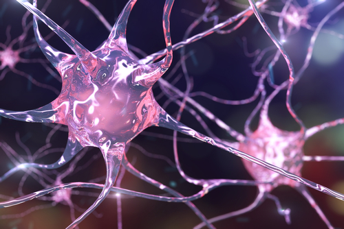
Manganese (Mn) is a trace metal that plays a key role in many cellular processes. It is essential in the production of neurotransmitters — chemical substances that allow communication between nerve cells — and in the regulation of nerve cells’ metabolism. However, high levels of Mn in the body are associated with neurotoxicity.
Levels of Mn change substantially in different regions of the brain throughout its development. However, it is still unclear if these regional differences could be linked to the fact that certain types of nerve cells may be more sensitive to higher levels of Mn than others at specific time-points during brain development.
Certain neurological disorders have been associated with alterations in brain Mn levels. It has been shown that human and mouse nerve cell precursors containing a genetic mutation associated with Huntington’s disease have limited access to Mn and are more resilient to its neurotoxic effects.
Investigators from Vanderbilt University and their collaborators now set out to explore the sensitivity of different types of neurons at different developmental stages, from patients with Huntington’s disease and healthy individuals (controls), to Mn neurotoxicity.
“We hypothesized that there would be differences in Mn sensitivity between lineages and developmental stages,” the researchers said.
The team used several lines of human-induced pluripotent stem cells (hiPSCs) — fully matured cells that can be reprogrammed back to a stem cell state, where they are able to grow into almost any type of cell — from patients and controls to generate neuroprogenitor cells (NPCs).
The NPCs then were cultured in a lab dish with different cocktails of growth factors to differentiate them into distinct types of neurons. Specifically, there were three different types: striatal neurons, which can be found in the striatum, a brain region involved in motor control; cortical neurons, which can be found in the cortex, or the outer layer of the brain; and midbrain dopaminergic neurons, which can be found in the substantia nigra, a brain region involved in the control of voluntary muscle movements.
The researchers then compared sensitivity to Mn neurotoxicity during each developmental time-point for each cell type between the two groups — those with and without Huntington’s.
Their findings revealed that striatal and cortical NPCs derived from Huntington’s patients were more resistant to high levels of Mn compared with those that had been obtained from individuals who did not have the disease. These results were similar to those seen in other studies.
Moreover, the investigators found that patient-derived hiPSCs were themselves more resistant to Mn neurotoxicity than their counterparts.
However, at intermediate stages of development, midbrain neurons that had been derived from patients became more sensitive to the toxic effects of Mn.
The researchers said the sensitivity of midbrain NPCs and mature cortical neurons to Mn neurotoxicity was similar in both groups.
Altogether, these findings suggest that the harmful effects of Mn can be influenced by the presence of genetic mutations associated with Huntington’s disease. That, in turn, depends on the particular developmental stages and neuronal cell types.
“In conclusion, our findings may provide insight into therapeutic strategies for diseases in which Mn has been shown to play a role such as HD [Huntington’s disease], especially through specific lineage-targeted interventions,” the researchers said.


Leave a comment
Fill in the required fields to post. Your email address will not be published.