
Enzymes Linked to Huntington’s Seen for 1st Time in Living Brain
Written by |
Scientists at the Martinos Center for Biomedical Imaging at Massachusetts General Hospital were able — for the first time — to track epigenetic factors, or non-genetic influences on gene activity, linked to Huntington’s disease in the brains of living humans.
The study, “Insights into neuroepigenetics through human histone deacetylase PET imaging,” published in the journal Science Translational Medicine, could advance the understanding of faulty brain processes in Huntington’s, as well as in other neurodegenerative and psychiatric diseases.
“The ability to image the epigenetic machinery in the human brain can provide a way to begin understanding interactions between genes and the environment,” Jacob Hooker, PhD, at the Martinos Center, and senior author of the report, said in a news release.
One of the most important processes for controlling which genes are active is the packaging of the long DNA strand into chromosomes. To form the densely packed structure of a chromosome, DNA is wrapped around proteins called histones. This wrapping is controlled using the addition or removal of specific molecules on the histones, referred to as epigenetic factors.
Acetyl is a common molecule used; the presence of an acetyl molecule is a signal allowing a protein to be formed from the gene, while removing the factor prevents the gene from being active. In Huntington’s disease, the enzymes responsible for removing the acetyl groups, called histone deacetylators or HDACs, are abnormally active, prompting scientists to develop drugs that block their activity.
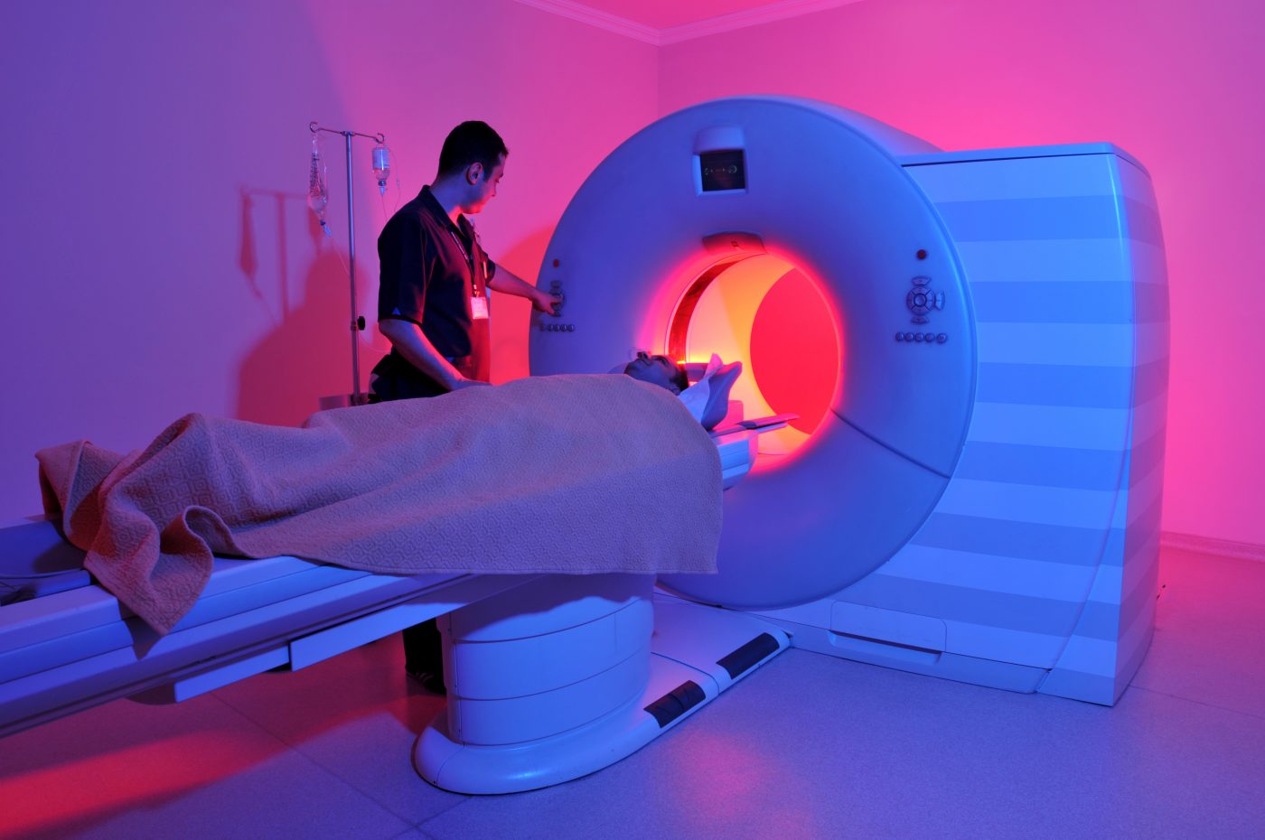
The researchers’ approach to analyzing epigenetics in living brains builds on emission tomography (PET), an imaging method that uses a weak radioactive trace molecule to study how tissues are working.
They developed a new tracer, dubbed Martinostat, that is able to visualize how many HDACs are produced in the brain. The tracer binds to the enzyme, and because it is radioactive, it lights up on a brain scan, allowing scientists to quantify enzyme levels. The level of radioactivity used in the tracer molecules is too low to damage the brain. Martinostat was tested in scans on eight healthy volunteers, and on human and animal brain tissues.
“HDAC dysregulation has been implicated in a growing number of brain diseases, so being able to study HDAC regulation both in the normal brain and through the progression of disease should help us better understand disease processes,” said Hooker, an associate professor of Radiology at Harvard Medical School.
Hooker said studies have begun on patients with several neurologic or psychiatric disorders.
“I believe Martinostat will help us understand the different ways these conditions are manifested and provide new insights into potential therapies,” he said.


Leave a comment
Fill in the required fields to post. Your email address will not be published.