Cerebellar Purkinje Cell Loss Linked to Huntington’s Motor Symptoms, Study Reports


The loss of a type of nerve cell called a Purkinje cell in a brain region known as the neocerebellum is associated with the presentation of motor symptoms in Huntington’s disease, a study finds.
The study, “Cerebellar degeneration correlates with motor symptoms in Huntington’s disease,” was published in the Annals of Neurology.
Although Huntington’s disease is caused by the disruption of a single gene known as huntingtin, patients show clear and considerable variability in motor symptoms. Recent studies have linked motor symptom variability in Huntington’s disease and the pattern of motor neuronal loss to two brain structures: the basal ganglia, involved in the control of voluntary motor movements, learning, cognition, and emotion, and the cerebral cortex, which plays a key role in memory, attention, awareness, language, and consciousness.
However, cell loss to these brain regions is not enough to explain the variability of motor symptoms observed in Huntington’s patients.
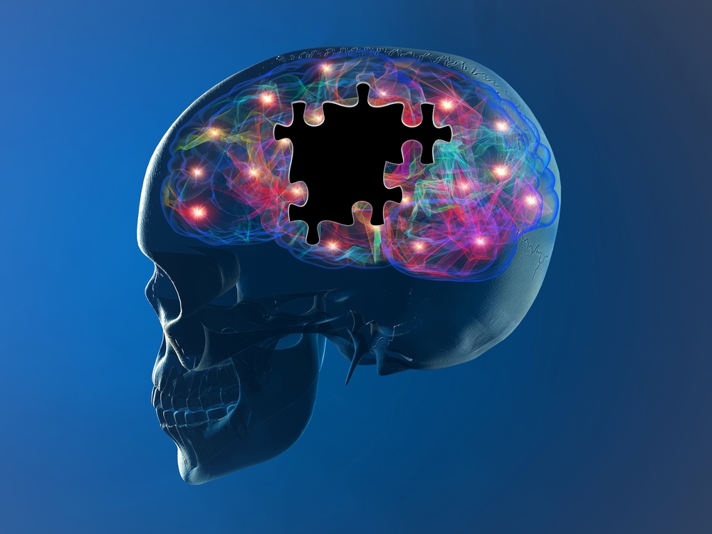
The cerebellum, a part of the brain located at the back of the skull, is known to play a crucial role in movement coordination, as well as posture and balance regulation, all of which can be affected by Huntington’s disease.
It is well-established that the neocerebellar region of the cerebellum plays a major role in the coordination of goal-directed voluntary movements, such as using a fork to eat or drinking a glass of water, which are especially affected in Huntington’s disease.
The loss of Purkinje cells — a major nerve cell type in the cerebellum — may also be a feature in Huntington’s disease cases with predominant motor symptom profiles. However, the role of the cerebellum and associated Purkinje cells in the development of Huntington’s disease was not unclear.
Researchers from the University of Auckland in New Zealand investigated whether Purkinje cell loss in the cerebellum was linked to Huntington’s disease and, if so, to motor symptom severity and presentation.
To do so, they compared post-mortem brain tissue from Huntington’s disease patients to brain tissue from controls — those without Huntington’s disease. The right cerebellum of 15 Huntington’s patients (10 men and five women, ages 35-66 years) and eight control patients (seven men and one woman, ages 41-68 years) were compared in the study.
Clinical data to assess Huntington’s disease symptom profiles were collected from clinical records and from family members for each case in the Neurological Foundation of New Zealand Human Brain Bank. These data were collected using an interview and questionnaire, and researchers administered them blindly, that is, without knowing if they were interviewing a Huntington’s patient.
Total clinical data for each case were reviewed independently by two neuropsychologists experienced with Huntington’s disease symptom assessments and were classified by the following:
- Motor: Individuals who had motor symptoms without the significant presence of mood symptoms during the symptomatic course of the disease.
- Mood: Individuals who mainly had mood disturbances during the symptomatic course of the disease but, to some extent, presented some very mild or late motor symptoms.
- Mixed mood-motor: those who had significant levels of both types of symptoms.
The team then matched this clinical data with very precisely mapped imaging data from the cerebellar samples examining Purkinje cell loss.
When comparing the results from all clinical and imaging data, the researchers did not observe a significant loss of Purkinje cells in the neocerebellum in Huntington’s disease samples compared with healthy controls.
However, they did find a link when they investigated the relationship between Purkinje cell loss and variable Huntington’s disease symptom profiles: Huntington’s motor cases showed a significant 43% loss of Purkinje cells in the neocerebellum compared with controls, while the mood sub-group showed no significant changes.
To make sure their findings were not influenced by other areas of the brain known to be affected by Huntington’s disease, the team examined whether the patients characterized as having motor issues were influenced by disease presentation in other brain structures, such as the striatum — a key component of the motor and reward systems.
The authors found that the degree of striatal pathology did not affect the results for disease symptoms. There was also no correlation between the total number of Purkinje cells with respect to CAG repeat length and age at death.
“This study … demonstrates, for the first time, a compelling relationship between Purkinje cell loss in the neocerebellum and motor symptoms in Huntington’s disease,” the researchers wrote.
The results show that multiple brain structures — the cerebellum, basal ganglia, and cerebral cortex — play an interactive role in the presentation of Huntington’s disease symptoms.
They also show how different each case of Huntington’s disease can be, which is likely why Huntington’s symptoms are so variable. These general conclusions may apply to other neurodegenerative diseases, such as Parkinson’s disease and Alzheimer’s disease.
“In conclusion, our stereological study detailing the loss of Purkinje cells in Huntington’s disease motor dominant cases demonstrates that the neocerebellum plays a significant role in the clinicopathological features of Huntington’s disease,” the researchers wrote.
“This study also supports the notion that along with the cerebral cortex and the basal ganglia, the cerebellum needs to be considered as a contributor to the symptom manifestations of Huntington’s disease,” they concluded.