Tracking Nerve Cells’ Signaling-protein Delivery Is Shedding Light on Huntington’s


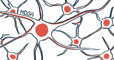
Nerve cells contain proteins that send signals to other cells and proteins that receive neighboring cells’ signals.
Researchers at Rensselaer Polytechnic Institute in Troy, New York, are trying to understand how the sending proteins end up on one side of a nerve cell’s membrane and the receiving proteins on the other side.
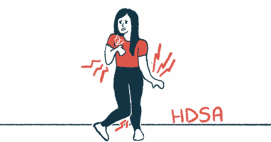
Understanding the process may help scientists identify the mechanisms that underlie the signaling-protein delivery errors that occur in neurodegenerative diseases like Huntington’s.
Nerve cells have special extensions that are responsible for sending and receiving signals. The sending extensions are called axons and the receiving ones dendrites.
The proteins in axons are different from those in dendrites. Scientists know that nerve cells produce both. What they haven’t understood is how the cells know to send signaling proteins to one side of the membrane and receiving proteins to the other side.
In Huntington’s and other neurodegenerative diseases, some of these proteins go to the wrong place. Understanding how protein sorting occurs will shed light on the mechanisms behind these diseases, scientists believe.
“This is a fundamental aspect of how the brain works, and no one really understands it,” Marvin Bentley, an assistant professor of biological sciences at Rensselaer Polytechnic Institute, said in a press release. “My goal is to answer basic scientific questions about this process,” said Bentley, who works in the institute’s Center for Biotechnology and Interdisciplinary Studies.
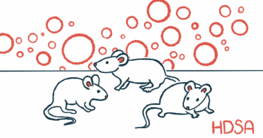
Bentley developed a series of techniques that allow researchers in laboratory settings to see how nerve cells transport proteins to their final membrane destination.
“Dr. Bentley’s work is making it possible to track proteins moving through a living neuron [nerve cell] in real time — a crucial component in the study of this basic neurological function,” said Curt Breneman, dean of the Rensselaer School of Science. “The information, and the insights derived from the live images he is producing, will advance the field of neuroscience and aid us in tackling crippling disorders. We are very pleased to welcome him as a colleague.”
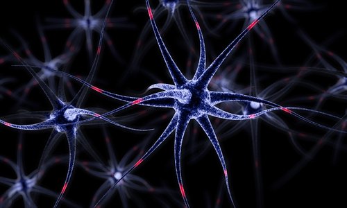
A nerve cell must use precise routing to deliver signaling proteins to its long axon and dendrite extensions. The key reason is that when a signaling protein ends up in the membrane, one side must be facing the interior of the cell and the other side facing outward.
Signaling-protein trafficking is constant. Cells must produce and move them to their final destination to replace proteins that wear out.
Nerve cells assemble the proteins in an area called the endoplasmic reticulum. From there, they travel to their ultimate location in small spheres, conveyed by a different class of protein — movement proteins.
Several steps regulate the journey. Bentley is particularly interested in the spheres and the movement proteins that carry them. Nerve cells have up to 20 different movement proteins.
By tagging different movement proteins with fluorescent tags and other labels, Bentley has been able to track them as they transport the spheres.
This has allowed researchers to observe which movement proteins are used, where they take the spheres, and how diseases such as Huntington’s may interfere with the spheres’ journey.
“Our goal is to figure out the very basics of how the cell works, how neurons work, and hopefully in the future it will be possible to correlate that back to these diseases and devise treatments,” Bentley said.