Autophagy Affects Protein Accumulation, Gene Activity in Huntington’s, Study Finds


Researchers have found a direct link between abnormal protein accumulation in nerve cells — a hallmark of several neurodegenerative diseases, such as Parkinson’s and Alzheimer’s — and the regulation of gene activity in Huntington’s disease.
According to researchers, both processes are influenced by autophagy, a mechanism used by cells to break down and recycle their own materials, that is frequently disrupted in several neurological disorders, including Huntington’s.
The findings of the study, “Huntingtin Aggregation Impairs Autophagy, Leading to Argonaute-2 Accumulation and Global MicroRNA Dysregulation,” were published in Cell Reports.
Huntington’s disease is characterized by the abnormal production and accumulation of a protein called huntingtin that is broken down into small pieces that stick together, forming toxic protein aggregates inside nerve cells that disrupt autophagy and, ultimately, lead to cell death.
The word autophagy is derived from the Greek words “auto” (self) and “phagy” (eating). It can be described as a natural cellular recycling process that deals with the destruction of damaged components of cells in the body to maintaining normal function.
Still, the implications of impaired autophagy for the function of nerve cells are not fully understood.
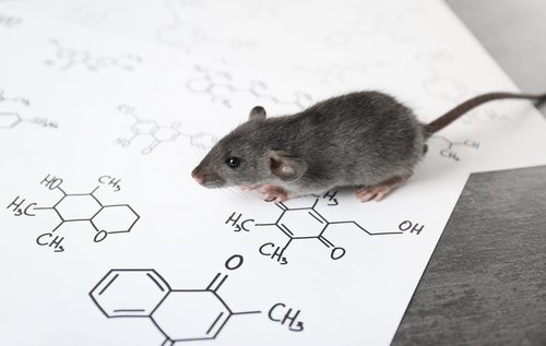
In this study, Lund University researchers in Sweden used a combination of cell culture techniques, mouse models of disease and post-mortem samples from patients with Huntington’s disease to investigate the impact of autophagy disruption in protein accumulation and gene expression in a context of disease.
Gene expression is the process by which information in a gene is synthesized to create a working product, like a protein.
“We know very little about why protein aggregates arise or in what way they are involved in the development of the disease. However, our study shows that expression of the mutated Huntingtin gene impairs the nerve cells’ ability to break down and recycle cellular material, which results in an accumulation of the protein AGO2,” Johan Jakobsson, PhD, a researcher in molecular neurogenetics and the study’s lead author, said in a press release.
Under normal conditions, Argonaute-2 (AGO2) — a protein involved in the regulation of microRNAs (miRNA, tiny RNA molecules that control the expression of multiple genes) — is usually degraded by autophagy. However, when autophagy is impaired due to the accumulation of huntingtin protein, AGO2 started to build up inside nerve cells.
As expected, when researchers artificially induced the activation of autophagy in the brains of animals with Huntington’s, AGO2 failed to accumulate inside nerve cells, confirming the involvement of autophagy in this process.
“It is worth noting that the number of HTT [huntingtin] inclusions was 3 times less [after autophagy activation], suggesting that autophagy activation … interferes with [disease] progression,” the authors wrote.
Moreover, scientists found that changes in autophagy also led to significant alterations in miRNA levels and activity.
“Our study indicates that changes in miRNA levels are an early sign of Huntington’s disease and are a result of changes in autophagy. It shows a direct link between protein aggregation and gene regulation,” said Karolina Pircs, PhD, a postdoc researcher on Jakobsson’s team.
Based on these findings, researchers are now convinced that novel autophagy-activating therapies should be developed to treat Huntington’s and other neurodegenerative diseases.
“If we can just find the right way to activate autophagy, we can perhaps effectively treat these diseases at an early stage. That is what we are currently working on in the lab,” Jakobsson added.