New Huntingtin Protein Strategy Significantly Improves Symptoms Over Time, Study Finds


A new therapeutic strategy based on a small part of the huntingtin protein — which in a mutated form is responsible for Huntington’s disease — shows potential to regulate the course of disease, according to a study using mice.
The study, “Improvement of BDNF signalling by P42 peptide in Huntington’s disease” was published in the journal Human Molecular Genetics.
Huntington’s disease is a neurodegenerative disorder caused by the expansion of an amino acid CAG sequence in the huntingtin (HTT) gene, leading to production of longer versions of the encoded huntingtin protein. (The CAG sequence is a very small stretch of DNA, that is repeated multiple times in a row. Healthy people normally have the CAG sequence repeated 10 to 35 times in their HTT genes. In people with Huntington’s disease, it may be repeated from 36 to more than 120 times.)
This abnormal protein is then cut into smaller toxic pieces that stick together and accumulate inside brain nerve cells, preventing their normal function or sometimes triggering cell death.
A team at University of Montpellier in France has identified a small part of the human huntingtin protein, called P42, that could prevent the clumping and accumulation of these toxic protein fragments.
To improve delivery of the small protein, researchers combined P42 with Medesis pharma’s proprietary Aonys technology. The innovative delivery system is based in a water-and-oil mixture that allows the delivery of the therapeutic agent to all cells of the body through oral or rectal ingestion.
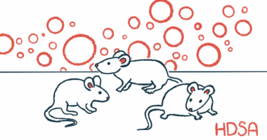
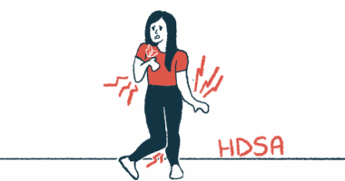
Treatment with P42 was found to significantly reduce brain atrophy in mice with established Huntington’s disease compared to animals treated with a placebo. The team also noticed significant improvements of about 18 percent in motor function two weeks after treatment initiation.
In addition, P42-treated animals showed reduced anxiety, and better learning and memory performance compared to placebo-treated mice.
A previous study showed that P42 significantly improved the behavioral manifestations of Huntington’s when given to mice that had the disease, but did not yet show symptoms. It also prevented changes in the brain and protein-fragment accumulation, which are associated with the disease.
The newer study further explored the potential of P42 therapy to treat Huntington’s disease by giving it to mice after symptom onset.
The findings suggest that the therapy used in the study could delay brain atrophy, significantly improve motor performance over time, and reduce anxiety in Huntington’s disease patients.
However, in contrast to the effect seen in pre-symptomatic mice, researchers found the treatment was unable to destroy the clumping of protein fragment aggregates in post-symptomatic animals.
“Although P42 prevented the formation of aggregates, it failed to destruct them once they are formed,” they wrote.
As such, the late effects of the treatment on brain cells’ fate “might involve other modes of action than its previously identified role on aggregation process,” they added.
In line with this theory, the team found that treatment with P42 significantly increased the levels of a molecule called brain-derived neurotrophic factor (BDNF), which is often reduced in Huntington’s patients and is necessary for nerve cell survival. The treatment also was associated with enhanced brain cell activity and plasticity.
Researchers believe “P42 offers an efficient therapeutic potential” by not only preventing aggregation of abnormal huntingtin fragments at early stages of the disease, but also by enhancing some natural functions of the normal version of the protein “at the different stages of the disease.”