Huntington’s Research to be Improved by New Mouse Brain Discovery


New data on an uncharted dark spot of the mouse brain map reveals how connections between brain cells form networks responsible for movement and motor learning — tasks that go awry in conditions such as Huntington’s disease.
The findings, discovered during a larger comprehensive project by scientists at the University of Southern California, will now allow researchers to focus on diseases such as Huntington’s, and target specific brain structures involved in the disease.
Led by Hong-Wei Dong, an associate professor of neurology at USC’s Mark and Mary Stevens Neuroimaging and Informatics Institute, the decade long Mouse Connectome Project is continually growing and ultimately aims to create the most complete atlas ever of a mammal brain – specifially the entire mouse brain.
New data about the project was recently included in the report “The mouse cortico-striatal projectome,” published in the journal Nature Neuroscience. The report included the previously uncharted brain region, the dorsal striatum, and its connections to the brain’s outer tissue layer called the cerebral cortex.
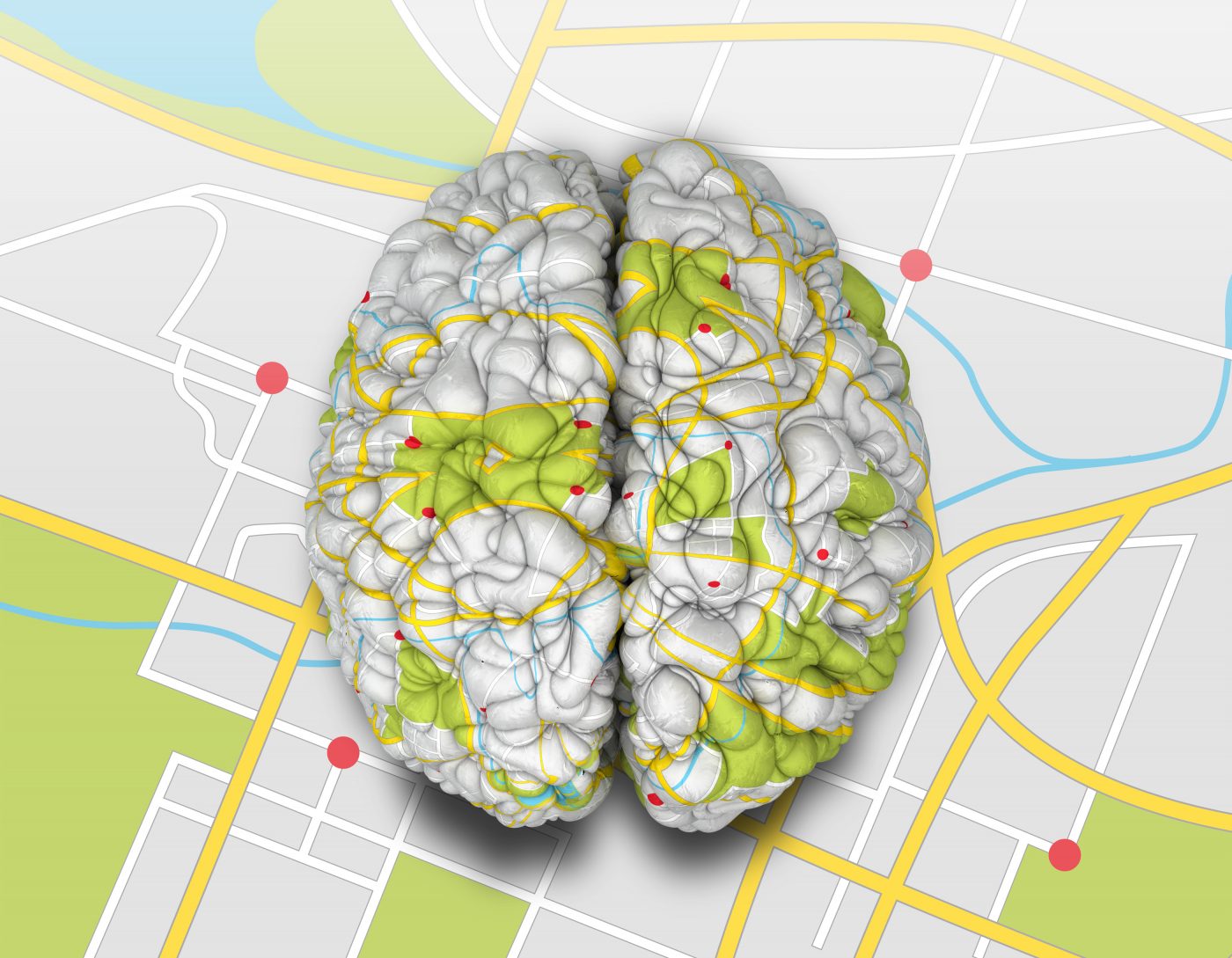
To achieve the impressive feat, Dong and his team used fluorescent molecules, which they injected into 150 different spots in the mouse brain. The procedure allowed them to trace how the molecules moved through the brain networks, which can be compared to a road map of highways connecting cities and smaller country roads. Like roads, the brain infrastructure needs to function smoothly in order to coordinate and execute behaviors.
In addition to Huntington’s disease, the brain region is of particular interest to scientists also studying Parkinson’s disease, obsessive-compulsive disorder, attention deficit hyperactivity disorder (ADHD), and other conditions that could be characterized by impulsive or uncontrollable movement.
“This study moves researchers to the next level to help them understand how the brain circuit is disrupted,” said Dong in a press release. “Previously the dorsal striatum was one huge thing. It’s almost like telling someone they should come visit you in California. Where should they go? We have really narrowed it down (to) I live in North Hollywood in this apartment building. That will help people in the future to really understand the pathways for diseases with specific symptoms.”
The team’s mapping efforts will allow researchers to focus on a certain part of the dorsal striatum. For example, for Huntington’s patients who exhibit early stages of experienced stuttering and abnormal eye movements, the ability to pinpoint and possible treat the exact network behind the actions would be a gigantic step forward.
The information extracted from mice allowed the team to divide the dorsal striatum into 29 parts that deal with various functions such as eye, mouth, and face movement. They also identified centers coordinating complex arm and leg movements.
“If you have one big structure, it’s very difficult to know which part is the problem area,” said Houri Hintiryan, lead author and assistant professor of research at the Laboratory of Neuro Imaging at the Keck School of Medicine of USC. “We’re providing a structural basis for studies seeking to understand which part of the brain does what.”
As always, the risk for slight differences between mouse and human brains exists, but like other types of research, mice models allow scientists to glean significant amounts of information that can be fine-tuned for human needs.
“Of course, humans and mice are different, but they are both mammals,” Dong said in a news release. “The biggest differences reside in high-level cognition. So we can use the organization of the mouse brain to understand how human brains are organized.”
Dong and his team will next hone in on the hippocampus, a brain region crucial for memory processing and emotion – and a region of major interest for Alzheimer’s researchers.
“With our brain map, researchers could look for circuitry-specific drug discovery,” Dong said. “Now, we provide a very clear map to help people do stem cell research. They know exactly where to put stem cells.”