Study Finds Novel Insight into ‘Ballooning Cell Death’ in Huntington’s Patients


A new Japanese study confirms that a phenomenon called “ballooning cell death” (BCD) occurs in patients with Huntington’s disease.
The study, “A novel form of necrosis, TRIAD, occurs in human Huntington’s disease,” appeared in the journal Acta Neuropathologica Communications.
Basically, three types of cell deaths occur in cells: apoptosis (cells program themselves to die after certain stimuli), necrosis (cells die suddenly due to particularly severe stimuli) and autophagy (cells start “eating” their own components for energy to survive, but if this process goes on too long, they die).
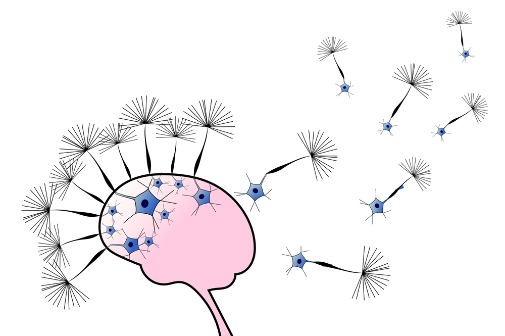
In late 2016, Hitoshi Hokazawa and his team at Tokyo Medical and Dental University observed that mutant huntingtin — the protein involved in Huntington’s disease — was associated with a novel mechanism called “ballooning cell death” (BCD). In this process, cells swell like a balloon until their membrane ruptures. BCD and necrosis are both triggered by reduced levels of two proteins that work together, TEAD and YAP, in a mechanism called TRIAD.
In the new study, Hokazawa used brains of human Huntington’s patients and mice with the disease to investigate the structural and biochemical aspects of this type of neuronal death.
They found that a protein called LATS1, which regulates the activation of TRIAD, was activated in brain tissue from patients and mice with Huntington’s, whereas Plk1 — which induces apoptosis rather than TRIAD — was inactivated. Moreover, levels of YAP/YAPdeltaC, which regulated TRIAD, in patients’ neurons fell.
Furthermore, researchers found that the endoplasmic reticulum, where most proteins are produced, was the main source of the ballooning in neurons from both humans and mice.
Together, these results provided information about how BCD and TRIAD unravel in the brain and identified several useful markers to determine the type of cell death involved in the pathology of Huntington’s.
“Our results showed activation of LATS1, suppression of Plk1, and decrease of YAP/YAPdeltaC,” researchers wrote. “[Microscopy] analysis also revealed typical morphological features of TRIAD. These data collectively supported that TRIAD occurs in human [Huntington’s disease] brains in vivo. In addition, biochemical and [microscopy] analyses revealed the chronological shift from early-phase to late-phase TRIAD changes, supporting the existence of TRIAD in the [Huntington’s disease] pathology in vivo.”
More than 30,000 Americans have Huntington’s, an inherited disorder that causes degeneration of brain cells in motor control regions of the brain, as well as other areas. HD typically begins between ages 30 and 50; there is currently no cure for the condition.