Huntington’s Pathology Seen in Songbirds’ Neuronal and Behavioral Response to HTT Mutation


New Huntington’s research sheds light on the link between the effects of the disease’s underlying mutation on brain structure and function, and consequent behavior anomalies. Studying the gene mutation’s expression in a precise brain area of songbirds, scientists observed how the mutation affects specific neurons and changes behavior, findings that could lead to the identification of a new therapeutic target.
The study, “Focal expression of mutant huntingtin in the songbird basal ganglia disrupts corticobasal ganglia networks and vocal sequences,” was published in the Proceedings of the National Academy of Sciences.
Huntington’s disease, caused by a mutation in the huntingtin (HTT) gene, greatly affects the basal ganglia, an area of the brain deeply involved in voluntary, sequential, and complex movements, learning, and cognition. Essentially, it helps to select motor gestures appropriate to a given behavioral context.
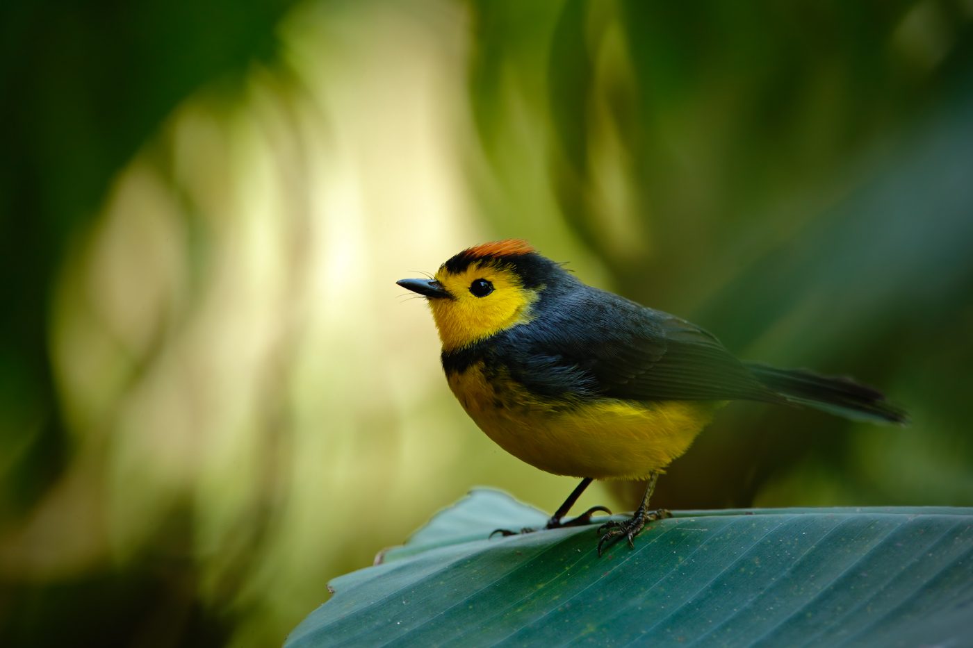
Although the genetic cause of Huntington’s is established, how the mutation affects brain structure and neuronal function so as to produce the behavioral and cognitive deficits, such as the disorganized movements observed in patients, is unclear. Songbirds are models to study the disease because the specific brain areas essential for learning and singing in these birds are highly developed. Moreover, each song is produced by a series of precise and repetitive movements in the vocal and respiratory muscles, so the reliability and repetitive nature of singing makes it possible to detect subtle changes as a response to a faulty gene.
Researchers expressed the genetic mutation that causes HD in the basal ganglia of songbirds, precisely in an area related to learning and singing. Within two months, they observed that these birds sang abnormal songs, and more songs, when compared to healthy birds. Behavioral changes included destabilization of syllable sequences and increased vocal activity, although the individual structure of each syllable remained intact. Such behavior was accompanied by the selective loss of medium spiny neurons. The main output neurons of the basal ganglia, the pallidal neurons, survived, but the timing of its synaptic activity became unreliable, suggesting a loss of neuronal complexity — which, if it could be replaced, may lead to the re-establishment of normal behavior. Importantly, the team observed that silencing the activity of this brain area partially rescued the disorganized vocals, leading to the stabilization of singing in a subset of birds. Apparently, no output from the basal ganglia is better than the wrong kind of output, Richard Mooney, the George Barth Geller Professor of Neurobiology in the Duke School of Medicine and the study’s senior author, said in a press release.
Researchers also believe that, unlike humans, songbirds may be able to generate new medium spiny neurons.
“If new neurons drive recovery, it may be that the songbird will provide a model for understanding how neural replacement in humans can be used to drive behavioral recovery in a range of neurodegenerative diseases,” Professor Mooney concluded.